X-Rays vs. Ultrasounds: How We Find What’s Going On Inside Your Pet
Sometimes our pets do things that make us go, “Really? Again?” One day, Persephone, a playful tabby, started vomiting. While occasional vomiting isn’t terribly unusual for a cat—especially one who loves to scarf her food like Perse—this time it was different. She wasn’t bringing up her food; instead, she vomited yellow bile, then stopped eating altogether. Her owners became very concerned.
Adding to the tension, one of her owners, an avid crafter, was busy working on a baby blanket for a friend ready to go into labor, making every moment of worry feel even more stressful.
Why We Start with X-Rays
Sometimes, cats like Persephone have ingested something that can’t be seen on X-rays. These items are called non-radiopaque, which means they don’t show up clearly on standard radiographs. Items like plastic, rubber, or certain fabrics often fall into this category.
Even so, we often start with X-rays because:
- They give a broad overview of the abdomen
- They can show changes in organ placement or gas patterns that hint at an obstruction
- They help us plan our next steps
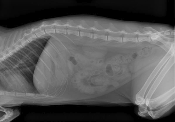
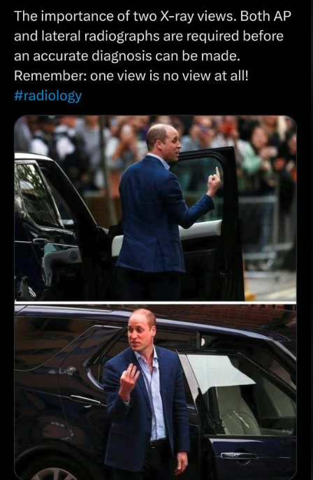
Think of X-rays like a treasure map, showing roughly where things might be, even if the “treasure” itself is invisible. To get the clearest picture, we usually take 2-3 X-ray views, from different angles, much like plotting coordinates on a map.
Most veterinary hospitals have 1-2 technicians in the X-ray room to help hold your pet safely for the best images. In some states, like New York, it is illegal for anyone other than the sedated patient to be in the X-ray room (link here). This means pets must be fully sedated to obtain images safely, which increases costs and carries some risk, though it protects staff. Current legislation is working toward similar safety standards in California.
When We Move to Ultrasounds
If X-rays don’t provide enough information, we move on to abdominal ultrasounds. Ultrasounds are perfect for:
- Locating soft tissue objects that X-rays can’t see
- Checking for blockages or obstructions
- Looking at organs in real-time, including the stomach and intestines
Ultrasounds use high-frequency sound waves to create live images. The probe is the most delicate instrument in the room, so it’s handled carefully. Sometimes a small patch of fur is shaved and very light sedation is used to keep pets calm and get the clearest images.

With Persephone, the ultrasound helped us pinpoint exactly where the non-radiopaque item was lodged, which is critical before considering surgery. It’s very hard to operate on a pet if we don’t know precisely where the object is stuck.
Why Both Imaging Methods Are Important
- X-rays: Provide a broad overview, detect bones, metal, or large objects, and show changes in organ placement
- Ultrasounds: Give a detailed look at soft tissues, locate non-radiopaque objects, and check for fluid or swelling
Often, your pet may need both tools to get a complete picture. Starting with X-rays and moving to ultrasound ensures we have the best information before making surgical decisions.
Who Reads the Ultrasound Matters
- Board-Certified Radiologist: Detects subtle or complex problems and provides a detailed report
- Primary Care Veterinarian: Can perform routine ultrasounds in-house and start treatment immediately
Your veterinarian will determine what’s best for your pet based on the situation.
? The Bottom Line: Imaging is essential to keeping pets safe and healthy. Whether it’s X-rays, ultrasounds, or both, these tools help us locate swallowed objects, detect blockages, and make informed treatment plans—so your mysterious kitty like Persephone can safely get back to eating normally and your family can breathe a sigh of relief.